Table 2 Descriptions and pictures of the different CARPs used for STL image quality evaluation
From: Stereolithography (STL) measurement rubric for the evaluation of craniomaxillofacial STLs
CARP | Description of CARP | Image of CARP |
|---|---|---|
1. Cranial sutures | The cranial sutures refer to a fibrous joint that holds bony plates together and only occurs in the cranium. |
Gray (1918) [14] |
2. Head of the mandible | The head of the mandible refers to the condyle, which presents an articular surface for articulation with the articular disk of the temporomandibular joint. |
https://media.cheggcdn.com/media/76c/76c5717c-0c6b-40ad-bee9-4ad78e2bc1fb/phpoRCXA2.png Chegg® Study (2021) |
3. Temporomandibular fossa separation | The temporomandibular fossa separation refers to the boundary between the temporomandibular fossa and the condylar head. |
Gray (1918) [14] |
4. Supraorbital foramina | The supraorbital foramina refer to the bilateral openings in the skull’s frontal bone located above the supraorbital margin of the orbits. |
Gray (1918) [14] |
5. Infraorbital foramina | The infraorbital foramina refer to the bilateral openings in the skull’s maxillary bone located below the infraorbital margin of the orbits. |
Gray (1918) [14] |
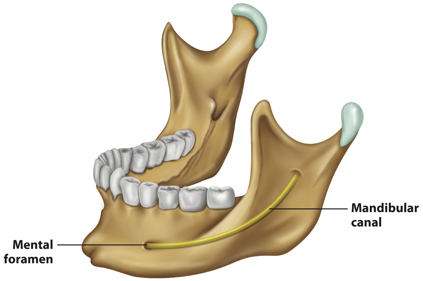
6. Mental foramina | The mental foramina refer to the two openings located on the mandible’s anterior surface. |
Gray (1918) [14] |
7. Teeth [15]a | The teeth refer to all the different types of teeth present in the mandible and maxilla. |
https://pubs.rsna.org/cms/10.1148/rg.307105026/asset/images/medium/105026fig04a.jpeg |
8. Mandibular canal | The mandibular canal refers to a canal within the mandible containing the inferior alveolar nerve, inferior alveolar artery and inferior alveolar vein. |
https://media.cheggcdn.com/media/76c/76c5717c-0c6b-40ad-bee9-4ad78e2bc1fb/phpoRCXA2.png Chegg® Study (2021) |


